Definition
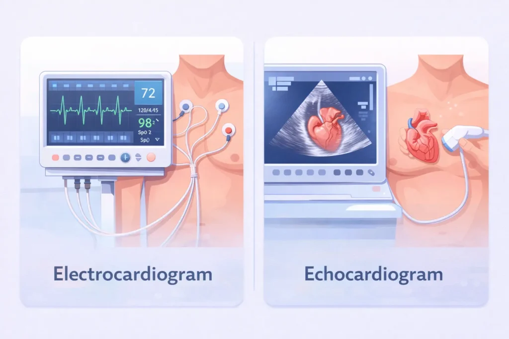
An electrocardiogram and an echocardiogram are both common heart tests, but they measure different aspects of heart health. An electrocardiogram records the electrical activity of the heart, while an echocardiogram uses ultrasound waves to create images of the heart’s structure and movement.
Heart health is essential for overall well being. When doctors suspect heart problems, they often recommend diagnostic tests to evaluate how the heart is functioning. Two of the most common heart tests are the electrocardiogram and the echocardiogram. Although their names sound similar, they serve different purposes and provide different types of information.
Many patients confuse these two tests because both are painless, non invasive, and frequently performed in hospitals and clinics. However, an electrocardiogram focuses on the electrical signals that control the heartbeat, while an echocardiogram creates detailed images of the heart’s chambers, valves, and blood flow using sound waves.
Understanding the differences between an electrocardiogram and an echocardiogram can help patients better understand their diagnosis and treatment plans. In this article, we will explain how each test works, their advantages and limitations, real world medical uses, global practices, and common misconceptions. By the end, you will clearly understand when each test is used and how it contributes to diagnosing heart conditions.
Quick Overview
Both tests examine the heart but measure different things.
| Feature | Electrocardiogram | Echocardiogram |
|---|---|---|
| Purpose | Measures heart electrical activity | Produces heart images using ultrasound |
| Technology | Electrical sensors on skin | Ultrasound sound waves |
| Detects | Heart rhythm abnormalities | Structural heart problems |
| Test Duration | 5 to 10 minutes | 20 to 40 minutes |
| Common Use | Detect arrhythmias and heart attacks | Evaluate valves, chambers, and pumping function |
Definition and Explanation
Electrocardiogram
An electrocardiogram is a diagnostic test that records the electrical signals produced by the heart. These signals control the heartbeat and help coordinate the contraction of heart muscles.
Small sensors called electrodes are placed on the chest, arms, and legs. These sensors detect electrical impulses as the heart beats. The signals are displayed as a graph on a monitor or printed on paper. Doctors analyze these wave patterns to detect abnormalities.
An electrocardiogram can reveal several conditions including irregular heartbeat, heart attack, and problems with the heart’s electrical conduction system.
The test is quick, painless, and usually takes only a few minutes.
Echocardiogram
An echocardiogram is an imaging test that uses ultrasound waves to create moving pictures of the heart. A small handheld device called a transducer sends sound waves into the chest. These waves bounce off the heart structures and return to the device, creating images on a screen.
These images allow doctors to observe:
- Heart chambers
- Heart valves
- Blood flow
- Heart muscle movement
Unlike an electrocardiogram, which measures electrical signals, an echocardiogram provides a visual view of the heart’s anatomy and mechanical function.
The test usually takes about 20 to 40 minutes and is also painless.
How Each Test Works
Electrocardiogram Process
- Electrodes are attached to the chest, arms, and legs.
- The patient lies still for several minutes.
- The machine records electrical signals of the heart.
- Results appear as wave patterns representing heartbeats.
Doctors analyze parts of the waveform to identify irregularities.
Echocardiogram Process
- Gel is applied to the chest.
- A transducer moves across the chest area.
- Ultrasound waves travel through the chest.
- The machine produces real time heart images.
These images help doctors evaluate heart structure and pumping efficiency.
Advantages and Disadvantages
Electrocardiogram Advantages
- Quick and simple procedure
- Detects abnormal heart rhythms
- Identifies heart attacks
- Widely available and inexpensive
- Non invasive and painless
Electrocardiogram Limitations
- Does not show heart structure
- May miss intermittent heart problems
- Cannot evaluate heart valves or muscle thickness
Echocardiogram Advantages
- Provides real time images of the heart
- Shows heart valves and chambers clearly
- Evaluates blood flow
- Detects structural abnormalities
Echocardiogram Limitations
- Takes longer than an electrocardiogram
- Requires specialized equipment
- Image quality may vary based on body structure
Types of Electrocardiograms
Doctors may use different types depending on the situation.
Resting Electrocardiogram
This is the most common test performed while the patient lies still.
Stress Electrocardiogram
This test records heart activity during physical exercise such as walking on a treadmill.
Holter Monitor
A portable electrocardiogram device worn for 24 to 48 hours to detect irregular rhythms that occur occasionally.
Types of Echocardiograms
Transthoracic Echocardiogram
This is the standard type where the ultrasound probe is placed on the chest.
Transesophageal Echocardiogram
The probe is inserted through the throat to obtain clearer images of the heart.
Stress Echocardiogram
Images of the heart are taken before and after exercise to assess blood flow during physical stress.
Real World Medical Examples
Doctors use both tests together to diagnose heart conditions.
1 Example
A patient with chest pain may first receive an electrocardiogram to check for a heart attack.
2 Example
If the electrocardiogram suggests abnormalities, an echocardiogram may be performed to examine the heart valves and muscle.
3 Example
A patient with shortness of breath may receive an echocardiogram to evaluate heart pumping function.
4 Example
Athletes sometimes undergo electrocardiogram screening to detect hidden heart rhythm disorders.
Regional and Global Usage
Heart diagnostic tests are widely used worldwide, but healthcare systems and technology availability influence how frequently each test is performed.
North America and Europe
Hospitals commonly use both electrocardiograms and echocardiograms as standard diagnostic tools. Emergency departments frequently perform electrocardiograms for patients with chest pain because it provides immediate results. Echocardiograms are widely used to evaluate heart structure and diagnose conditions such as heart valve disease and heart failure.
Asia
Countries such as Japan and South Korea have highly advanced cardiac imaging systems. Preventive heart screening programs often include electrocardiograms. Echocardiograms are commonly used for early detection of congenital heart disease and cardiomyopathy.
Middle East
Modern hospitals in many Middle Eastern countries use advanced echocardiography systems for diagnosing structural heart conditions. Electrocardiograms are widely used in emergency care and routine checkups.
Africa and Developing Regions
In some regions, electrocardiograms are more accessible because they are cheaper and easier to perform. Echocardiograms may be limited to larger hospitals due to equipment costs.
Global Trends
Telemedicine and portable devices are expanding access to heart testing. Portable electrocardiogram monitors and handheld echocardiography devices are increasingly used in remote locations.
Common Mistakes and Misconceptions
Many people misunderstand the difference between these tests.
Mistake 1
Believing both tests are the same.
Correction
They evaluate different aspects of heart function.
Mistake 2
Thinking an electrocardiogram shows heart images.
Correction
It records electrical activity, not visual images.
Mistake 3
Assuming an echocardiogram detects heart rhythm problems.
Correction
It focuses mainly on structure and blood flow.
Mistake 4
Believing one test replaces the other.
Correction
Doctors often use both tests together for complete diagnosis.
Related Concepts and Comparisons
Electrocardiogram vs Echocardiogram vs Stress Test
| Test | Purpose | Technology |
|---|---|---|
| Electrocardiogram | Detect heart rhythm abnormalities | Electrical sensors |
| Echocardiogram | Evaluate heart structure | Ultrasound imaging |
| Stress Test | Assess heart response to exercise | ECG monitoring during exercise |
Exercises for Learning
Exercise 1
Identify whether the following situations require an electrocardiogram or echocardiogram.
1 A doctor wants to check irregular heartbeat.
2 A doctor needs to evaluate heart valves.
3 A patient experiences chest pain in the emergency room.
4 A doctor wants to measure heart pumping strength.
Answers
1 Electrocardiogram
2 Echocardiogram
3 Electrocardiogram
4 Echocardiogram
Exercise 2
Match the feature to the correct test.
| Feature | Electrocardiogram | Echocardiogram |
|---|---|---|
| Records electrical signals | Yes | No |
| Creates heart images | No | Yes |
| Detects arrhythmias | Yes | No |
| Evaluates heart valves | No | Yes |
FAQs
What is the main difference between electrocardiogram and echocardiogram?
An electrocardiogram records the electrical activity of the heart, while an echocardiogram produces ultrasound images showing the heart’s structure and movement.
Which test is better for diagnosing heart problems?
Both tests are important. Electrocardiograms detect rhythm problems, while echocardiograms reveal structural abnormalities.
Is an electrocardiogram painful?
No. Electrocardiograms are painless and involve attaching electrodes to the skin.
Is an echocardiogram safe?
Yes. Echocardiograms use ultrasound waves and do not expose patients to radiation.
How long does an electrocardiogram take?
Most electrocardiograms take between five and ten minutes.
How long does an echocardiogram take?
An echocardiogram typically takes twenty to forty minutes.
Can an electrocardiogram detect heart attacks?
Yes. Electrocardiograms are often used in emergency departments to identify signs of a heart attack.
Can an echocardiogram show heart valve problems?
Yes. Echocardiograms are one of the best tests for evaluating heart valve structure and function.
Why do doctors sometimes order both tests?
Using both tests gives doctors a complete understanding of heart electrical activity and structural function.
Do these tests require special preparation?
Usually no preparation is required, although patients may be asked to avoid heavy exercise before certain tests.
Conclusion
Electrocardiograms and echocardiograms are two of the most important diagnostic tools in cardiology. While their names are similar, they evaluate different aspects of heart health. An electrocardiogram measures the electrical signals that control heart rhythm, while an echocardiogram uses ultrasound technology to produce detailed images of heart structures.
Both tests are non invasive, painless, and widely used in medical practice. Electrocardiograms are especially useful for detecting arrhythmias and heart attacks, while echocardiograms help doctors evaluate heart valves, chambers, and pumping ability.
Doctors often use both tests together because they provide complementary information. Understanding the difference between electrocardiogram and echocardiogram helps patients feel more confident about their diagnosis and treatment.
Maintaining heart health through regular checkups, healthy lifestyle choices, and early diagnostic testing can significantly reduce the risk of serious cardiovascular disease.
Discover More Related Articles:
- Titanium vs Tungsten: Which Metal Is Best for Jewelry and Rings? in 2026
- Alternately vs Alternatively: Which One Should You Use in Writing in 2026
- Moles vs Freckles: What Your Skin Is Really Saying in 2026

David Thompson is the grammar expert behind TalkNexs.com. He believes that learning English grammar doesn’t have to be boring or complicated. Through practical examples and easy explanations, David helps readers understand confusing grammar rules and use them correctly in daily communication. His goal is to make grammar simple, clear and enjoyable for everyone.